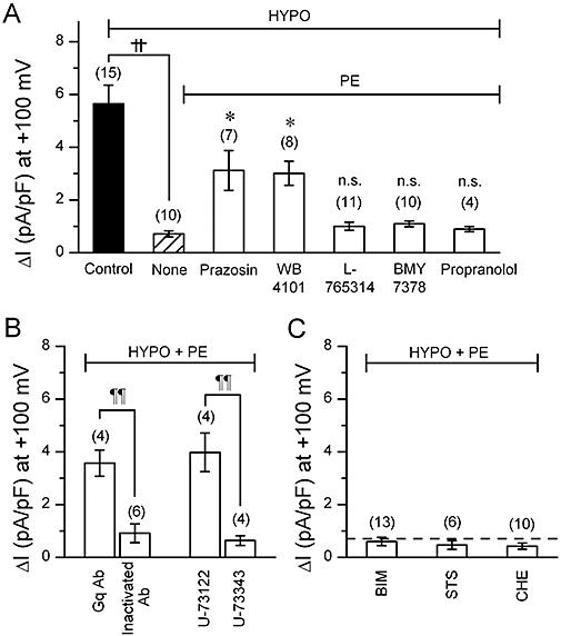
Figure 5.

Volume-regulated anion channel (VRAC) current observed in cells with various α1 adrenoceptor–related compounds. (A)–(C), mean density of hypotonicity-induced (difference) currents measured at +100 mV under various conditions. Phenylephrine (PE) concentration was 100 µM throughout. (A) shows the selective effect of α1A-adrenoceptor antagonists on the PE-induced VRAC inhibition. The cells were exposed simply to hypotonic solution (HYPO) (Control) or to PE-containing HYPO. In the latter case, the cells were pretreated with 5 µM prazosin, 2 µM WB-4101, 50 nM L-765314, 30 nM BMY-7378 or 1 µM propranolol for 10 min under isotonic condition, and then exposed to PE-containing HYPO in the continuous presence of these antagonists. Some cells were given no antagonist (None). ††, significantly different with P < 0.01. *significantly larger than None with P < 0.05. n.s., not significantly different from None. (B) shows effects of anti-Gq antibody (Gq Ab) or a phosphoinositide-specific phospholipase C (PLC) inhibitor, U-73122, on PE-induced VRAC inhibition. For Gq Ab experiments, Gq Ab or heat-inactivated Gq Ab was added to the pipette solution at a 1:100 dilution. For the experiments of PLC inhibitor, the procedure of drug application was similar to that adopted in the experiments shown in A, that is, it included a pre-treatment phase of 10 min. U-73122 (5 µM) or its inactive analogue, U-73343 (5 µM) was used. ¶¶, significantly different with P < 0.01. (C) shows the lack of effect of PKC inhibitors on PE-induced VRAC inhibition. The procedure of drug application was similar to the above. Bisindoylmaleimide (BIM; 100 nM), staurosporine (STS; 10 nM) or chelerythrine (CHE; 10 µM) was used. Number in parentheses indicates number of the cells examined. Dashed line indicates the average magnitude of VRAC current with PE, derived from the data shown as None in (A).
