
FIG. 1.
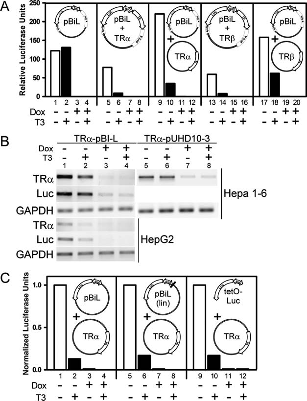
Regulation of pBi-L by doxycycline and thyroid hormone (T3). (A) From left to right, the pBi-L plasmid itself (pBi-L), the same vector with a TRα1 insert, the pBi-L plasmid together with a pSG5-TRα1 expression vector, the pBi-L vector with a TRβ1 insert, or the pBi-L plasmid together with a pSG5-TRβ1 expression vector were transiently transfected into Hepa 1-6 cells using the lipofection procedure previously described (1). Transfections also included a pCH110 β-galactosidase construct as an internal normalization control (1). Cells were maintained at 37°C in Dulbecco-modified Eagles medium containing 10% hormone-depleted fetal bovine serum. After 24 hours the transfection medium was replaced with medium containing doxycycline, T3, or both, as indicated below each panel. After an additional 24 hours the cells were harvested and relative luciferase activity was calculated (1) and is presented. (B) The overall transfection protocol in panel A was repeated using either the TRα1-pBi-L construct described above, a related (TRα-pUHD10-3) construct based on a similar plasmid lacking the luciferase gene (pUHD10-3), and either Hepa 1-6 cells or HepG2 cells as indicated. After harvesting the cells, mRNA was isolated and quantified for levels of TRα1, luciferase, and GAPDH using a reverse transcription PCR protocol (4). (C) Alternatively, the pBi-L vector, a linearized version of the pBi-L vector (lin), or a fragment of the same construct limited to the luciferase-bidirectional promoter region (tetO-Luc) were tested together with the TRa expression vector in the same transfection/luciferase assay as described in panel A.