
FIG. 3.
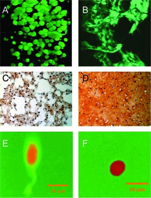
(A, B) Human mesenchymal stem cells (hMSCs) encapsulated in a photopolymerized hydrolytically biodegradable PEG hydrogel and cultured in osteogenic medium. After encapsulation and when limited degradation has occurred, hMSCs adopt a spherical morphology (A). With sufficient degradation, hMSCs are able to migrate and form cell–cell junctions and adopt a more osteogenic-like phenotype (B). Reproduced with permission from Cushing et al.53 (C, D) Engineered cartilage after 2 (C) and 6 (D) weeks in vitro culture (glycosaminoglycans stained red). Chondrocytes were encapsulated in a biodegradable PEG-co-PVA hydrogel exhibiting bimodal degradation. Reprinted with permission from Martens et al.56 (E, F) Fibroblasts encapsulated in an enzymatically degradable PEG hydrogel. When crosslinks are degraded, fluorescence is emitted at the degradation site enabling spatial visualization of the degrading gel. After 7 days postencapsulation, cell-mediated degradation was observed only in the direction of the extended process (E); however, when treated with an MMP inhibitor, no cell-mediated degradation was observed (F). Reproduced with permission from Lee et al.64 Color images available online at www.liebertpub.com/ten.