
Figure 6.
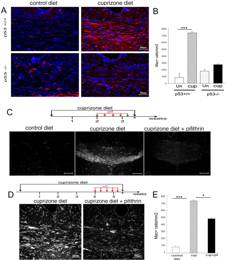
Pifithrin-α treatment of cuprizone-fed mice reduces recruitment of microglial cells to the corpus callosum even if initiated after oligodendrocyte apoptosis has occurred. A, Confocal image of coronal brain sections of wild-type (p53+/+) and knock-out (p53−/−) siblings fed either a control diet or a 0.2% cuprizone diet for 4 weeks and stained with Mac1 antibody (red) to identify microglial cells and with DAPI (blue) as nuclear counterstain. B, Bar graphs indicate the relative number of Mac1+ cells detected in the dorsal corpus callosum of p53+/+ and p53−/− mice receiving either control (Un) or 0.2% cuprizone (cup) diet. n = 3 animals per condition; ***p < 0.001. C, Schematic representation of the duration of the early pifithrin-α treatment (red arrows) in mice fed a cuprizone diet for 3.5 weeks (black arrows). The asterisk denotes the time point of analysis when the animals were killed and the brains were processed for immunohistochemistry (top). Immunohistochemistry of coronal brain sections of the medial corpus callosum at the level of the dorsal fornix, stained with antibodies specific for the microglial marker Mac1 (bottom), is shown. Note the presence of immunoreactive cells only in the cuprizone-fed mice but not in mice on a control diet or in mice treated with pifithrin-α during the first 3 weeks of cuprizone diet. Scale bars, 80 μm. D, Schematic representation of the duration of the late pifithrin-α treatment (red arrows) in mice fed a cuprizone diet for 5.5 weeks (black arrows). The asterisk denotes the time point of analysis when the animals were killed and the brains were processed for immunohistochemistry. Immunohistochemistry of coronal brain sections of the medial corpus callosum at the level of the dorsal fornix, of mice treated as described in C, is shown. Sections were stained with antibodies specific for the microglial marker Mac1. E, Bar graph representation of Mac1+ cells in the dorsal corpus callosum of animals that received either a control diet or a 0.2% cuprizone diet for 5.5 weeks in the absence (cup) or presence of systemic pifithrin-α (cup+pif) administration during the fourth week. n = 3 for each treatment; ***p < 0.001; *p < 0.05. Error bars indicate SEM. pif, Pifithrin-α.