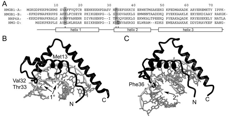
Figure 1.
HMGB sequences and structures. (A) Sequence alignment of HMGB1 boxes A and B, NHP6A, and HMGD. The sequences are aligned and numbered according to the HMGD structure with the alpha helices depicted by rectangles. Residues known to intercalate the DNA are shaded in grey. (B) The ribbon diagram shows the crystal structure of wildtype HMGD bound to DNA (PDB ID 1qrv), with the intercalation sites indicated by arrows and the intercalating residues labeled. (C) The ribbon diagram shows the crystal structure of HMGB1 box A bound to cisplatin-modified DNA (PDB ID 1ckt), with the intercalation site indicated by an arrow and the intercalating residue labeled.

