
FIG. 4.
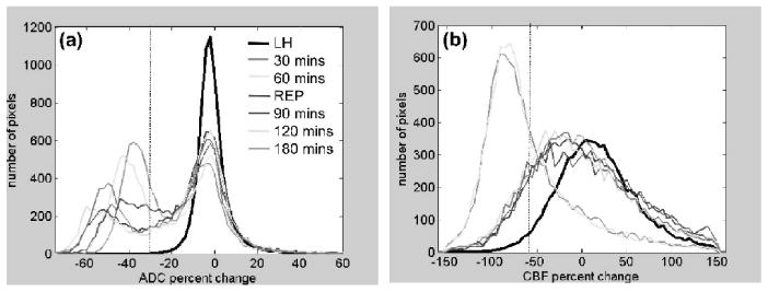
(a) ADC and (b) CBF projection profiles from the scatterplots in Fig. 3 (n = 6) as a function of time (30 and 60 minutes, pre-reperfusion; 70, 90, 120, and 180 minute, post-reperfusion). Pixel distributions were plotted for the entire left hemisphere (for the five time points, excluding 70 minutes, and divided by five for displaying on the same scale). Pixel distributions were plotted for the entire right hemisphere for each time point. The ADC distribution was bimodal but the CBF was uni-modal. Superimposed on these distributions are the vertical dotted lines indicating TTC-derived ADC (- 30%) or CBF (- 57%) viability thresholds as previously determined (Shen et al., 2003).