
Fig. 1.
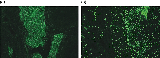
Serum immunoglobulin (Ig)A endomysium antibodies (EMA) and nuclear fluorescence reactivity (NFR) antibody patterns (Olympus microscope, 400×). (a) Serum IgA EMA-positive. A fluorescence feature of EMA is a honeycomb-like staining pattern along muscolaris mucosae (endomysium) in cryostat sections of the third distal portion of monkey oesophagus. Specifically, EMA react against the collagenous matrix of type 3 connective tissue surrounding the smooth muscle fibres of the primate oesophagus. (b) Serum IgA NFR-positive. Fluorescence feature of NFR is a dot-shaped staining pattern on nuclei of muscular and epithelial cells in the same sections.