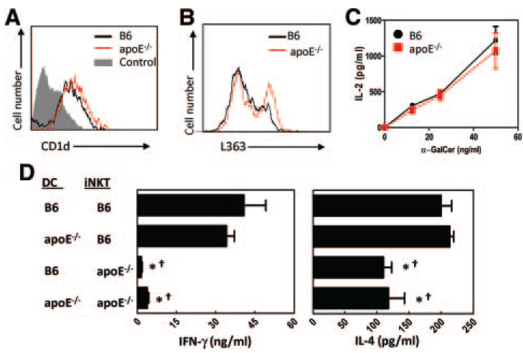
Figure 4.
DCs from apoE−/− mice retain the ability to activate iNKT cells. A, CD11c+ splenic DC expression of CD1d as determined by flow cytometry. B, Splenocytes from B6 and apoE−/− mice were incubated 18 hours with 50 ng/mL GGC, then stained with 1 µg/mL L363 monoclonal antibody to detect surface CD1d:α-GalCer complexes. Flow cytometry analyses were performed by gating on CD11c+ cells. Shown are representative histograms of 2 experiments using 3 mice per group. C, CD11c+ DCs were purified from spleens of B6 and apoE−/− mice, loaded with 50 ng/mL α-GalCer for 30 minutes, then incubated with the iNKT cell hybridoma DN32.D3 for 48 hours. Activation of the hybridoma, as measured by IL-2 production in culture supernatants, was determined by ELISA. α-GalCer alone (with no DC) did not result in IL-2 production. D, Purified splenic DCs were incubated with 50 ng/mL α-GalCer for 30 minutes, then added to purified iNKT cells and cocultured for 72 hours. IFN-γ (left panel) and IL-4 (right panel) were determined by ELISA. *P<0.05 when compared with B6 DCs cultured with B6 iNKT cells, and †P<0.05 when compared with apoE−/− DCs cultured with B6 iNKT cells, as determined by 1-way ANOVA.

