Figure 5.
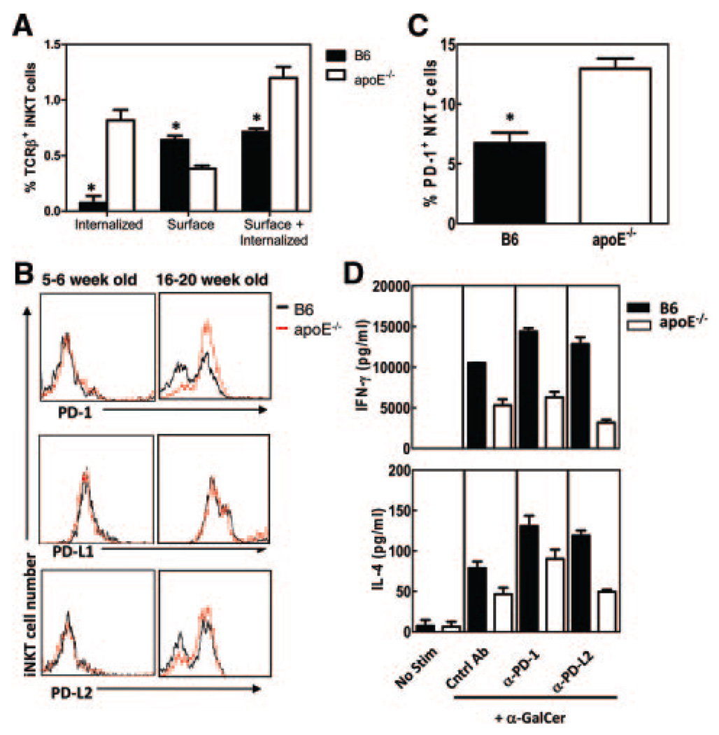
iNKT cells from apoE−/− mice have increased PD-1 expression. A, iNKT cells from 16- to 20-week-old B6 and apoE−/− mice were stained for internalized and surface TCRβ expression and analyzed by flow cytometry. Internalized TCRβ was determined by subtracting surface TCRβ from surface+internalized TCRβ. Shown are representative histograms from 3 mice per group. B, Ex vivo PD-1, PD-L1, and PD-L2 expression on iNKT cells from 5- to 6-week-old (left panels) and 16- to 20-week-old (right panels) B6 and apoE−/− mice. Shown are representative histograms from 3 experiments. C, Percent PD-1+ iNKT cells ex vivo in 16- to 20-week-old B6 and apoE−/− mice. D, Splenocytes were cultured in vitro in the presence or absence of α-GalCer, with 50 µg/mL of control immunoglobulin, anti-PD-1, or anti-PD-L2 antibodies. After 72 hours, IFN-γ (top panel) and IL-4 (bottom panel) were determined by ELISA in culture supernatants. *P<0.05 as determined by Student’s t test. No stim indicates no stimulation.

