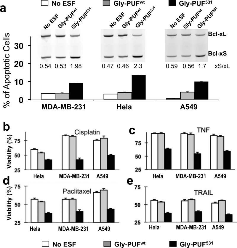
Fig. 5. ESFs affect Bcl-x splicing in multiple cancer cells.
(a) Splicing modulation of Bcl-x in three cancer cell lines infected with lentivirus expressing Gly-PUF531, control ESF (Gly-PUFwt) or GFP (as mock infection). Bcl-x splicing isoforms were detected by RT-PCR, with Bcl-xS% being quantified and listed (inset). For reasons we are not completely clear, the lentivirus infected Hela cells have elevated basal level of Bcl-xS. Propidium iodide stained cells were analyzed with flow cytometry to determine the fraction of dead cells. (b) Effect of ESFs on cisplatin sensitivity of different cancer cells. Cells were infected with lentivirus to express the same designer ESF and controls, and cisplatin was added to a final concentration of 5 μM at 72 hours after infection. Cell viability was measured with the WST-1 assay 24 hours after drug treatment. All treatments were repeated at least twice, and the means with error bars representing the standard deviation (n=3) from representative experiments are plotted. White bars represent cells of mock infection, grey bars represent control Gly-PUFwt infection and black bars represent Gly-PUF531 infection. (c, d and e) Effect of ESFs on the sensitivities to paclitaxel, TNF-alpha and TRAIL in different cancer cell lines. Experimental conditions are the same as described for panel b except final concentrations of 10 nM paclitaxel (c), 20 ng/ml TNF-alpha (d), or 100 ng/ml TRAIL (e) were used. The significant differences (P<0.05, judged by paired T-test) of cell viabilities were observed for all drug treatments between the Gly-PUF531 and Gly-PUFwt infected cells.

