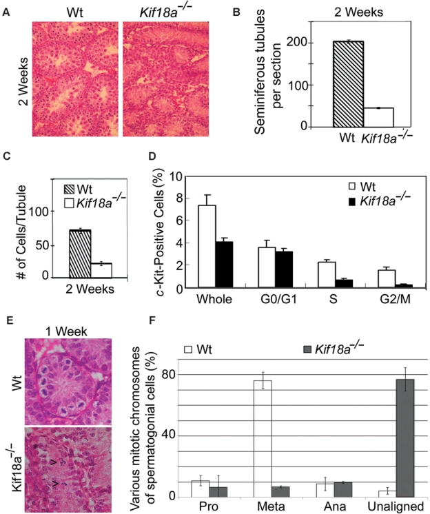
Figure 2.

Mitotic defects in Kif18a−/− mouse testes. (A) Representative sections of wt and Kif18a−/− at week 2 were stained with H&E. (B) Seminiferous tubules were counted from 6 random fields of each wt or Kif18a−/− testis. The data were summarized from 3 independent mice. (C) Cells in each seminiferous wt or Kif18a−/− tubule were counted from 6 random fields of each testis. The data were summarized from 3 independent mice. (D) The c-Kit-positive cells isolated from wt or Kif18a−/− testes were subjected to the DNA content analysis by flow cytometry. Representative results are shown. (E) Representative wt and Kif18a−/− seminiferous tubules at week 1 are presented. Arrowheads denote mitotic cells with unaligned chromosomes. (F) Mitotic cells in week 1 wt or Kif18a−/− seminiferous tubules were examined under microscope. Cells of various mitotic stages or with unaligned chromosomes were quantified.
