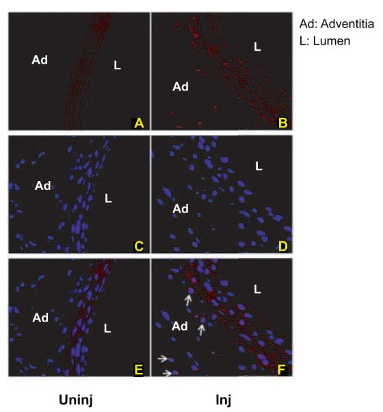
Figure 2.

Nuclear ETS-1 expression is increased in injured carotid arteries. Shown are ETS-1 antibody immunofluorescence–stained sections at 24 hours postinjury (Inj) and uninjured carotid arteries (uninj). Perfusion-fixed and sectioned arteries were immunostained using primary antibodies against ETS-1. The secondary antibody was Texas Red conjugated (A and B). The sections were mounted with 4′,6-diamidino-2-phenylindole mounting medium (C and D). Merging of the 2 pictures with Adobe Photoshop shows nuclear colocalization of ETS-1 (E and F, arrows).
