Figure 6.
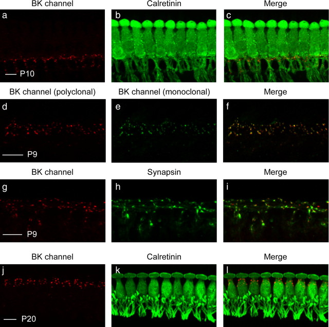
Immunostaining reveals transient expression of BK channels in the region of the MOC efferent–IHC synapses. a, b, Cochlear preparations from P10 mice were immunostained with a rabbit polyclonal antibody against the BK channel (a) and a mouse monoclonal antibody against calretinin (b), a cytoplasmic marker of IHCs as well as the afferent fibers contacting IHCs. c, BK channel immunoreactivity is visible as small puncta in the region below IHCs. d, e, To verify the specificity of BK channel immunolabeling, cochlear preparations from similarly aged mice were immunostained with both a rabbit polyclonal (APC-021; d) and a mouse monoclonal (L6/23; e) antibody against the BK channel. f, The colocalized immunoreactivity suggests that both antibodies are specifically detecting the BK channel. g, h, To determine whether BK-positive puncta below IHCs are associated with efferent terminals, cochlear preparations were immunostained with a rabbit polyclonal antibody against the BK channel (g) and a goat polyclonal antibody against synapsin (h), a cytoplasmic marker of efferent presynapses. i, BK-positive puncta indeed correspond with synapsin-positive efferent terminals. j, k, Finally, cochlear preparations from P20 mice were immunostained with a rabbit polyclonal antibody against the BK channel (j) and a mouse monoclonal antibody against calretinin (k). l, At this age, BK channel immunoreactivity is no longer visible as small puncta below IHCs but is instead restricted to larger puncta surrounding the neck of IHCs. Scale bars, 10 μm.

