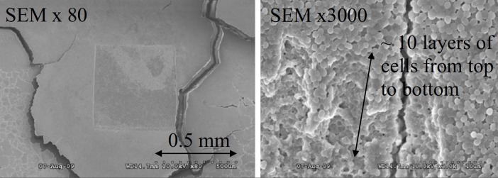
FIG. 4.

(Left) SEM image of an area of ablated on the surface of thick biofilm ( thick). Laser was focused to a spot diameter of and ablation was done by rastering laser beam across the surface with spacing between shots. Magnification was 80×. (Right) Top right corner of the ablated region shown on the left image. Magnification was 3000×. The depth penetration was estimated by counting layers of cells from the top to the bottom, as the diameter of an individual S. epidermidis cell is .
