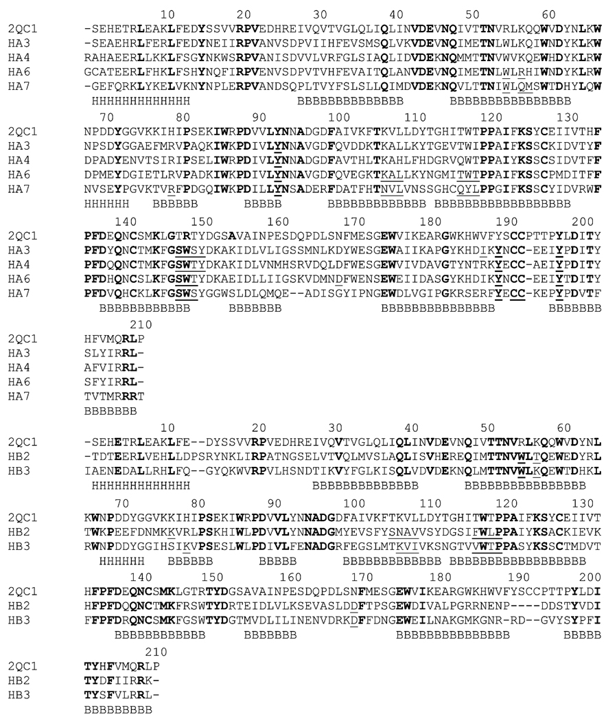
Fig. 1.
Multiple sequence alignment of the ECDs of the mouse α1 with human nAChR monomers. The alignment was divided into subunit 1 monomers (top, α3, α4, α6, and α7) and subunit 2 monomers (bottom, β2, and β3). Identical residues are shown in bold, while residues involved in ligand binding are shown in underline. Secondary structure elements are shown under the sequences: H= a-helix, B=b-strand.

