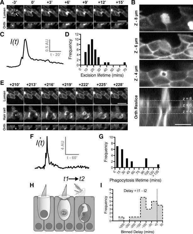
Figure 2.
Supporting cells drive epithelial repair in the avian utricle using two distinct mechanisms. Transgenic utricles (E17–E19) expressing β-actin-EGFP were time-lapse imaged live in the presence of 1 mm streptomycin to induce hair cell death. A, E, A time-lapse sequence of hair cell removal and epithelial repair after at least 12 h of streptomycin treatment. Panels depict the exact same hair cell at different times during the repair process. In this mosaic, only supporting cells (stars) were expressing β-actin-EGFP. A nonexpressing hair cell was present in the center of the mosaic (arrow). A, Activity was initially restricted to the epithelial lumen where supporting cells formed an actin cable (0′) that invaded into the hair cell (+3′) and excised the stereocilia bundle (+6′). Junction complexes reformed after removal of the stereocilia bundle (+15′). B, Higher-resolution laser-scanning confocal image of a different hair cell shows how the actin cable constricts beneath the cuticular plate to eject the stereocilia bundle. C, Changes in fluorescence intensity of supporting cell β-actin-EGFP measured at the luminal surface during stereocilia excision. D, Distribution of supporting cell activity during stereocilia excision (n = 36 events). E, After stereocilia excision (shown in A) supporting cells engulfed the remaining hair cell soma (star) within a phagosome highlighted by β-actin-EGFP. F, Supporting cell activity during phagocytosis was heterogeneous and was best modeled as a spiking phenomenon. Frame-by-frame differential fluorescence intensity was used to detect suprathreshold activity. The initial burst of activity represents formation of the phagosome, followed by an extended period where the cell is internalized. G, Distribution of supporting cell activity during phagocytosis (n = 22 events). H, Schematic of the hair cell removal process. I, Distribution of delay (t1 − t2) between onset of stereocilia excision (t1) and phagocytosis (t2). The majority of stereocilia excision events occurred in advance of hair cell phagocytosis. Scale bars: A, E, 10 μm; B, 5 μm. Times are expressed in minutes (′).

