
Figure 4.
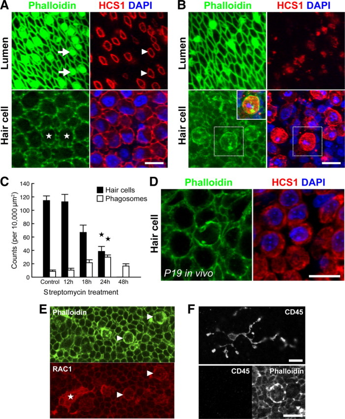
Supporting cell phagosomes are present in aminoglycoside-treated utricles, both in vitro and in vivo. Wild-type chick utricles (E21) were cultured with 1 mm streptomycin sulfate in vitro to induce hair cell toxicity. Paraformaldehyde fixed samples were labeled with anti-HCS-1, phalloidin, and DAPI, and then visualized using confocal microscopy. A, Normal organization of the sensory epithelium after 24 h in vitro. Phalloidin highlights the stereocilia bundles at the epithelial lumen (arrows) and anti-HCS-1 labels the hair cell pericuticular junction (arrowheads). Within the epithelium, hair cell somas (stars) are packed densely together with HCS-1 labeling the basolateral membranes. B, After 24 h exposure to 1 mm streptomycin, there is significant hair cell loss. Few stereocilia bundles are visible and this correlates with expansion of supporting cells at the epithelial lumen. At the luminal surface, the HCS-1-positive rings associated with the hair cell pericuticular regions are no longer visible. At the level of the hair cell nuclei, there are HCS-1-labeled cells containing pyknotic DNA. F-actin accumulates in phagosomes containing HCS-1 and pyknotic DNA (inset, overlay of boxed regions). C, Quantification of hair cell and phagosome density in utricles incubated with streptomycin for varying durations reveals an inverse relationship. A significant change is observed at 24 h (compared to sham-operated controls; n = 5 utricles per group;  p < 0.001). D, Phagosomes were also detected in P19 utricles after systemic administration of streptomycin in vivo. E, F, Phagosomes colocalize with phagocytic, but not macrophages markers. Phagosomes (arrowheads) and a macrophage (star) both label with anti-RAC1 (CED-10) (E). However, phagosomes do not label with anti-CD45 (F, bottom), unlike macrophages (F, top). Scale bars: 10 μm. Data are expressed as mean ± SEM.