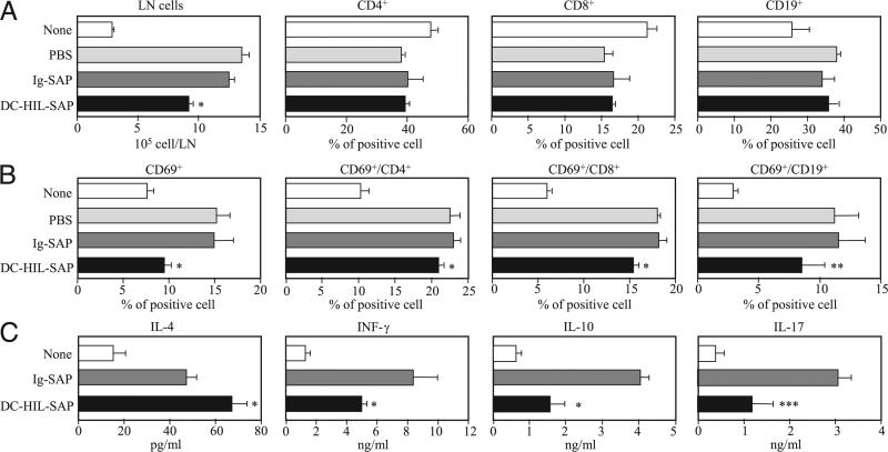
FIGURE 5.
Immunological phenotypes of DLN cells from mice treated with SAP conjugates. A and B, Sensitized mice (n = 3) were i.v. injected with PBS, Ig-SAP, or DC-HIL-SAP (each 40 nM) 3 h prechallenge. Two days postchallenge with Ox, DLNs procured from treated mice were counted and expressed as cell number per DLN. Frequency (%) of CD4+, CD8+, and CD19+ B cells was measured by flow cytometry (A). DLN cells also were examined for expression of CD69 on all LN cells, CD4+, CD8+, or CD19+ cells, and frequency (%) was calculated (B). C, DLN cells from similarly treated mice (n = 3) were cultured for 2 d in the presence of anti-CD3 and anti-CD28 Ab and assayed for production of IL-4, IFN-γ, IL-10, and IL-17. As control, DLN cells from unsensitized mice (None) were also analyzed in the same manner. SD was derived from experimental values for three mice. Statistical significance is denoted by asterisks (*p < 0.001; ** p = 0.14; *** p = 0.03) as compared with frequency in LNs of mice treated with control Ig-SAP. Data shown are representative of three separate experiments.

