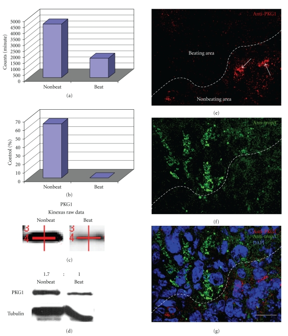
Figure 1.
Graphs show raw and normalized data taken directly from the Kinexus protein kinase screen. (a) Kinexus Inc. (http://www.kinexus.ca/) uses a highly sensitive imaging system with a 16-bit camera (Bio-Rad Fluor-S Max Multi-Imager) in combination with quantitation software (Bio-Rad Quantity One) to quantify and analyze the chemiluminescent samples. The resulting trace quantity for each band scanned at the maximum scan time is termed the raw data. As the relationship between scan time and band intensity is linear over the quantifiable range of the signal intensity, the raw data from the scans are normalized to 60 seconds (counts per minute—C.P.M.) for uniformity. (b) After normalization, data are converted to percentages by subtracting the control (Nonbeating) normalized C.P.M. from the experimental (Beating) normalized C.P.M., followed by dividing the difference by the control (Nonbeating) normalized C.P.M. and multiplying by 100. (c) The actual PKG1 bands from the Kinexus report show a stronger signal in beating areas when compared to nonbeating areas. The red lines in (c) are generated and used by the computer to find the correct bands. (d) Followup Western blots confirmed the Kinexus data. A ratio of PKG1 protein levels was calculated by normalizing to tubulin showing that there is 1.7X (69%) more PKG1 in nonbeating areas versus beating areas. (e)–(g) Confocal microscopy of EBs showed higher levels of PKG1 in nonbeating areas, represented by anticardiac troponin (e)–(f), when compared to nonbeating areas. Arrows point to PKG1 within cells negative for cardiac troponin. Scale bar in G =100 micrometer.

