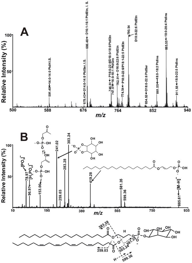
Figure 6.
MALDI-TOF MS analysis of phospholipids in the negative ion mode using 9- aminoacridine as matrix. A) A MALDI mass spectrum of negatively charged phospholipids from murine myocardium was obtained by examining an aliquot of a Bligh and Dyer extract of myocardium by MALDI MS in the negative ion mode using a 4800 MALDI-TOF/TOF Analyzer as described in the “Experimental Section”. The prefix “D” and “P” stand for diacyl (i.e., phosphatidyl-) and alkenyl-acyl (plasmenyl-) species, respectively. ‘‘IS’’ denotes internal standard. B) A MALDI tandem mass spectrum of fragment ions obtained from 18:0–20:4 PtdIns present in mouse heart lipid extracts acquired on a 4800 MALDI-TOF/TOF Analyzer in the negative ion mode. The tandem mass spectrum was recorded on a 4800 MALDI-TOF/TOF Analyzer in the negative ion mode using 9-aminoacridine as matrix using CID with the metastable suppressor on and the timed ion selector enabled. The voltages of source 1, collision cell and collision cell offset were 8.0 kV, 7.0 kV and −0.035 kV, respectively. The tandem MS spectrum was obtained by averaging 2000 consecutive laser shots (50 shots per subspectra with 40 total subspectra).

