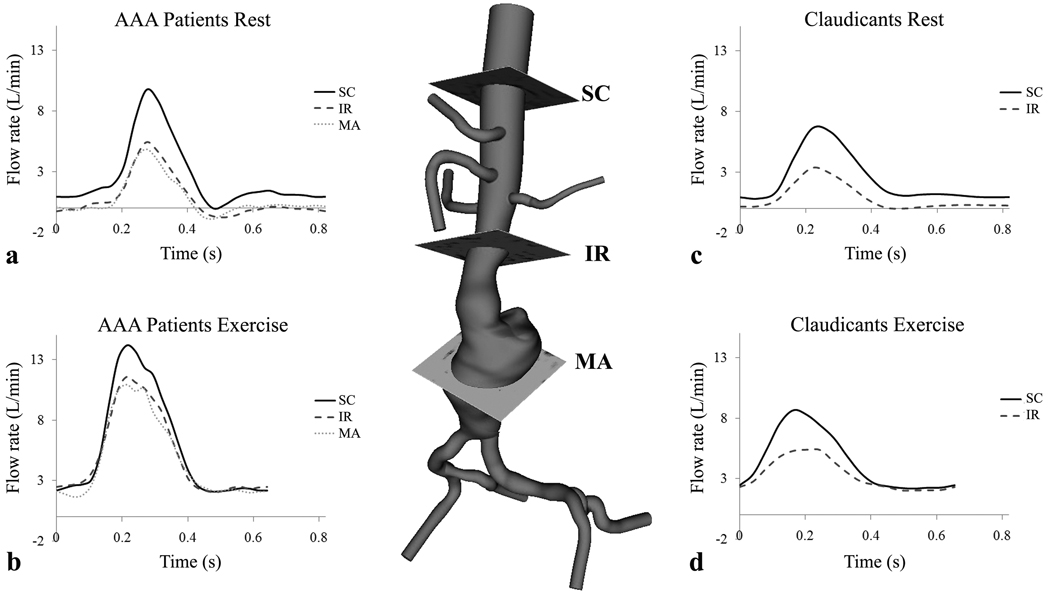
Figure 2.
A three-dimensional model of an AAA and imaging slices for supraceliac (SC), infrarenal (IR), and mid-aneurysm (MA) positions. During rest, AAA participants had retrograde flow at each anatomic location (a), and claudicants had retrograde flow at the infrarenal level only during diastole (c). During exercise, the peak systolic blood flow rate increases, diastole shortens, and retrograde flow is eliminated for AAA participants (b) and claudicants (d). Note that at rest, AAA participants exhibited a triphasic waveform (a) compared to a biphasic waveform for claudicants (c).

