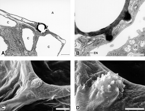
Fig. 1.
Transmission (A and B) and scanning (C and D) electron micrographs of puffball spores deposited on alveolar surfaces. Notice that the spore particle is totally covered by the surface lining layer and submerged. The epithelium was indented even by the particle's spiny protrusions (at these locations, the particle is separated from the capillary by 100 nm). A, alveolar lumen; C, capillary; EN, endothelial cell; EP, epithelial (type 1) cell; LC, leukocyte; LL, osmophilic lining layer material. Scale bars = 2 (A and D), 0.5 (B), or 5 μm (C). [From Geiser et al. (18)].

