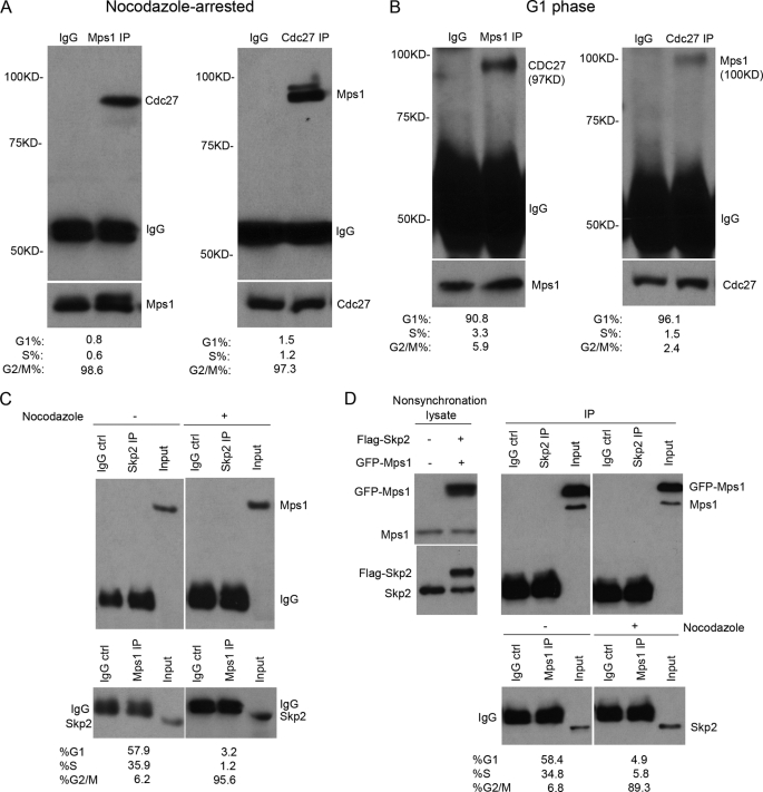
FIGURE 2.
Mps1 associates with the APC-c, but not the SCFSkp2, complex in vivo. Co-IP assays of Mps1 and Cdc27 in nocodazole-arrested (A) and G1-synchronized 293T cells (B). Note that a longer film exposure was required to detect Mps1-Cdc27 complexes in G1-synchronized cells due to the lower levels of Mps1 present. Cells were incubated with nocodazole (40 ng/ml) for 16 h prior to arrest in G2/M. A double thymidine protocol was used for G1 synchronization. The protein expression levels are shown at the bottom. During the last 6 h before harvesting, cells were treated with MG132 (25 μm). C, co-IP assays of endogenous Mps1 and Skp2 (component of SCF) performed with asynchronous and nocodazole-arrested 293T cell lysates. Mps1 did not co-precipitate with endogenous Skp2. D, GFP-Mps1 and FLAG-Skp2 proteins do not associate with each other in vivo following transient expression in 293T cells. Co-IP assays were performed using anti-Mp1 and anti-Skp2 antibodies. IPs using normal mouse IgG were performed as negative controls in all experiments. Cell cycle distribution for asynchronous, G1-synchronized, and nocodazole-arrested cells was assessed by flow cytometry.

