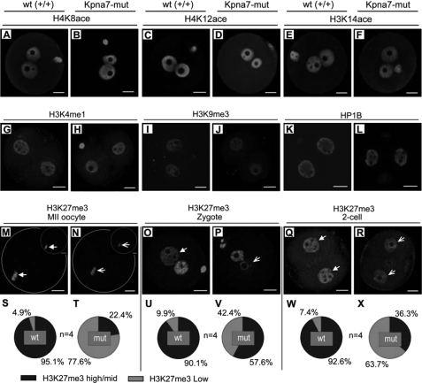
FIGURE 8.
Epigenetic analysis of oocytes, zygotes, and two-cell embryos isolated from Kpna7 mutation mice. A–F, histone acetylation analysis. H4K8ace (A and B), H4K12ace (C and D), and H3K14ace (E and F) levels in zygotes of Kpna7 mutation mice and wild type mice are similar. G–L, H3K4me1, H3K9me3, and HP1B staining. H3K4me1 (G and H), H3K9me3 (I and J), and HP1B (K and L) staining levels in late two-cell embryos of Kpna7 mutation mice and wild type mice are similar. M–R, H3K27me3 staining in MII oocytes, zygotes, and parthenogenesis two-cell embryos. H3K27me3 levels are down-regulated in MII oocytes (M, N, S, and T) and zygotes (O, P, U, and V) of Kpna7 mutation mice. DAPI and H3K27me3 double staining are shown in top right of M and N to show their colocalization. H3K27me3 levels are down-regulated in parthenogenesis two-cell embryos (Q, R, W, and X) but not in normally fertilized two-cell embryos (data not shown) of Kpna7 mutation mice. (Solid arrows indicate the high levels of staining in wild type group, and open arrows indicate the low levels of staining in Kpna7 mutation group; bars, 20 μm).

