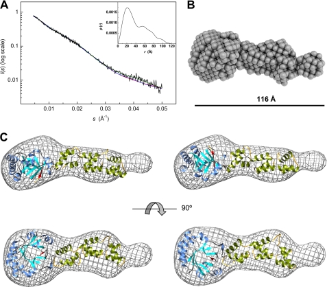
FIGURE 7.
SAXS-based modeling of Cpl-7 overall structure. A, experimental SAXS profile of Cpl-7 (black line) and theoretical spectra calculated from the ab initio bead model (green line) in B, and the high resolution models in C. The inset shows the pair-distance distribution function, P(r), generated by GNOM with a Dmax of 115 Å (data collected at 4 °C in Pi buffer). B, ab initio bead model of Cpl-7 derived from SAXS data using DALAI_GA (50). C, molecular envelope (grid representation) of Cpl-7 bead model with the best fits of the three-dimensional models built for the CM and the CWBR manually docked inside (the antiparallel β8-strand of the CM is colored in red). Red and blue lines in A are the theoretical SAXS profile derived for models a (left-hand model) and b (right-hand model), respectively, using CRYSOL (54).

