FIGURE 7.
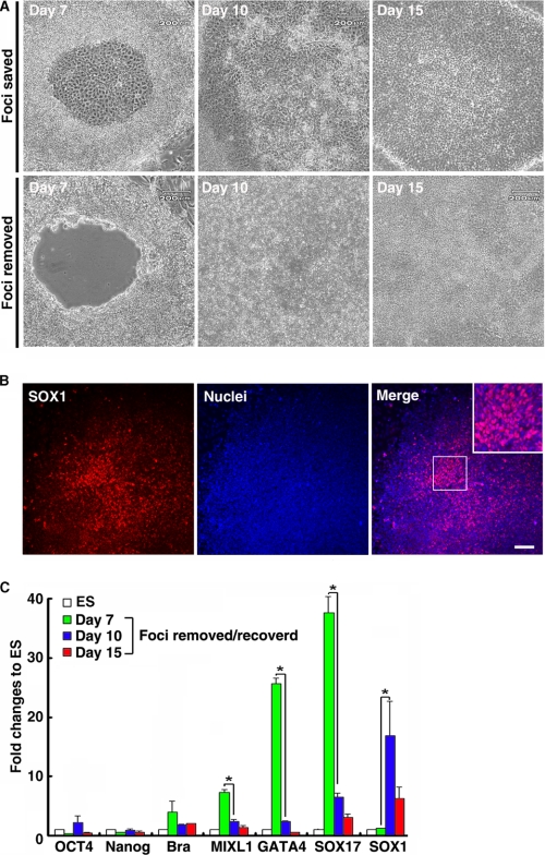
Removal of the central foci of monkey ESC colonies from IVDS2 disrupted hepatic endoderm differentiation under hESdF-primed culture conditions. A, the differentiated cells in the central area of ESC colonies at early IVDS2 (day 7) were excised and removed with a glass micropipette. The central area of an ESC colony was repopulated by cells grown from peripheral areas 3 days (day 10) after cell removal. B, ICC for SOX1 on ESC colonies 3 days after cell removal, showing that the central area of ESC colonies was repopulated with SOX1-expressing cells. Scale bar, 25 μm. C, QRT-PCR analysis of total RNA isolated from foci removed IVDS2 (day 7), regenerated cells at 3 (day 10) and 8 days (day 15) after central area removal using primers specific for Oct4, Nanog, Brachyury, MIXL1, GATA4, SOX17, and SOX1. The data correspond to the means and standard deviations of triplicate experiments. *, p < 0.05 by Student's t test.

