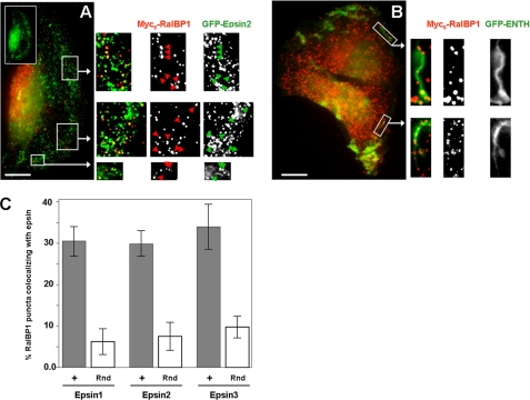
FIGURE 2.
GFP-epsin/ENTH domain partially co-localizes with RalBP1 at the plasma membrane in ruffles and puncta. HT1080 cells co-transfected with Myc6-RalBP1 and GFP-epsin2 (A) or GFP-ENTH (B) were extracted with saponin, fixed, and immunolabeled. Arrows highlight some areas of co-localization. The inset shows polarization of GFP-epsin2 toward the leading edge. Scale bar: 10 microns. C, percentage of RalBP1 puncta colocalizing with each of the epsins was quantified. Approximately 200 puncta per cell were counted in 6 different cells. To estimate the probability of random colocalization, the image corresponding to the RalBP1 channel was rotated 180° and random signal overlap was assessed (Rnd, open bars).

