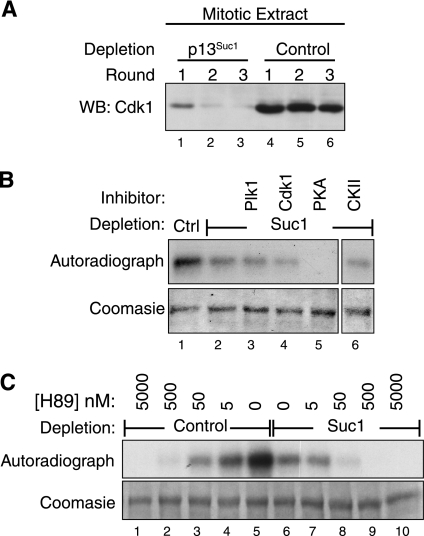
FIGURE 4.
PKA and CDK1 contribute to CUX1 phosphorylation in mitosis. A, mitotic cells lysates from nocodazole-arrested T98G cells were prepared in nondenaturing conditions. CDK1 was depleted from the extract using three successive incubations with p13Suc1-agarose beads or beads alone as a control. Immunoblotting was done using CDK1 antibodies. Lysates corresponding to lanes 3 and 6 were used as a source of kinase in subsequent experiments. WB, Western blot. B, His-CUX1(612–1328) was purified from bacteria and used as a substrate in kinase assays using, as a source of kinases, either control or CDK1-depleted lysates (lanes 1 and 2, respectively). Reactions were done in parallel in the presence of inhibitors against PLK1 (500 nm BI2536), CDK1 (2.5 μm CGP74514A), PKA (20 μm H89), or CKII (60 μm 4,5,6,7-tetrabromobenzotriazole), as indicated. C, kinase reactions were performed as in B, in the presence of various concentrations of the PKA inhibitor H89. WB, Western blot.

