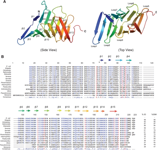
FIGURE 3.
Structure of the periplasmic domain of LptC. A, a ribbon diagram of a single His6-LptC(24–191) molecule at 2.2-Å resolution. The structure of the periplasmic domain of LptC is composed of a series of 15 antiparallel β-strands that wind back along the path of the preceding peptide stretch throughout the length of the protein, resembling the structure of LptA. B, an alignment of the sequences of selected LptC homologues; predicted secondary structure features are identified. Residue numbering corresponds to LptC from Neisseria meningitidis (without gaps). Alignment was performed with MultAlin (59). Residues with high sequence identity or similarity are shown as colors, and the overall identity (%ID) and similarity (%SIM) are reported. Non-conserved residues are shown as black letters.

