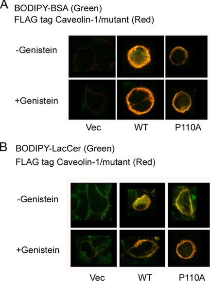
FIGURE 2.
BODIPY-BSA and BODIPY-LacCer internalization in HEK 293 cells transfected with either caveolin-1 or the P110A mutant. A, top row, the cells were incubated with 50 μg of BODIPY-BSA for 30 min at 10 °C, washed, and warmed for 5 min at 37 °C before back-exchange. The cells were fixed with PFA and permeabilized with 0.1% Triton X-100 prior to indirect immunofluorescence using an antibody directed against the FLAG tag and an Alexa 647 secondary antibody. BODIPY-BSA was observed at green wavelengths, and Alexa 647 (FLAG) was observed at far red wavelengths (see “Experimental Procedures”). The figure shows the merged images. Bottom row, the cells were pretreated with 80 μm genistein, an inhibitor of caveola-mediated endocytosis, prior to incubation with fluorescent markers (2 h at 37 °C) and during the above mentioned steps. Columns show examples of different cells. B, top row, the cells were incubated with 2 μm BODIPY-LacCer·BSA for 30 min at 10 °C, washed, and warmed for 5 min at 37 °C before back-exchange. The cells were treated as for A. Bottom row, the cells were pretreated with 80 μm genistein as for A. There was background green fluorescence in the plasma membrane of control cells (the vector-transfected cells). Pictures show the midportion of cells using the confocal fluorescence microscopy. Columns show examples of different cells. Vec, vector.

