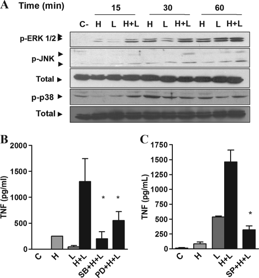
FIGURE 4.
Heme anticipates and increases the activation of MAPKs induced by LPS. A, macrophages from C57Bl/6 mice were stimulated with heme (100 μm) and/or LPS (0.1 ng/ml) in the time intervals indicated. Cell extracts were submitted to SDS-PAGE. ERK1/2, JNK, and p38 phosphorylations were detected by immunoblotting. Detection of nonphosphorylated ERK2 was used as loading control. The figures are representatives of three different experiments with similar results. B and C, production of TNF was evaluated in peritoneal macrophages treated with SB203580 (10 μm), PD98059 (30 μm) (B), or SP600125 (30 μm) (C) for 30 min before the stimuli with heme (100 μm) and LPS (0.1 ng/ml). The supernatants were collected 4 h after the stimuli. Results represent means ± S.E. for TNF determination and are representative of two different experiments. *, p < 0.05. H, heme; L, LPS; SB, SB203580; PD, PD98059; SP, SP600125.

