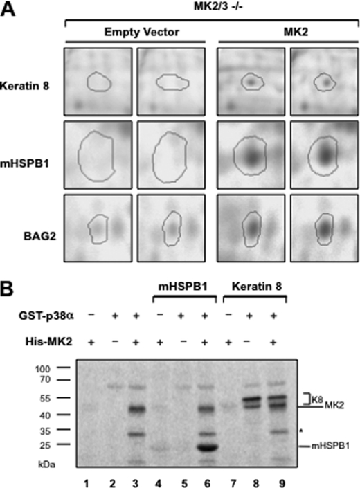
FIGURE 1.
Reduced K8 phosphorylation in MK2/3-double deficient MEFs. A, MK2/3-deficient cells stably transduced with empty vector and MK2 expression vector in duplicates were stimulated with 10 μg/ml anisomycin for 30 min. Cell lysates were analyzed by two-dimensional PAGE and stained with Pro-Q Diamond phosphoprotein gel stain. The spots for K8, mHSPB1, and BAG2 with reduced staining in the gels of MK2/3-deficient cell lysates are shown. B, K8 and mHSPB1 were subjected in parallel to in vitro radioactive kinase assays with purified His-MK2 (lanes 4 and 7), GST-p38 (lanes 5 and 8), or with both (lanes 6 and 9). Reactions were separated on 7.5–22.5% gradient SDS-PAGE and analyzed by phosphorimaging.

