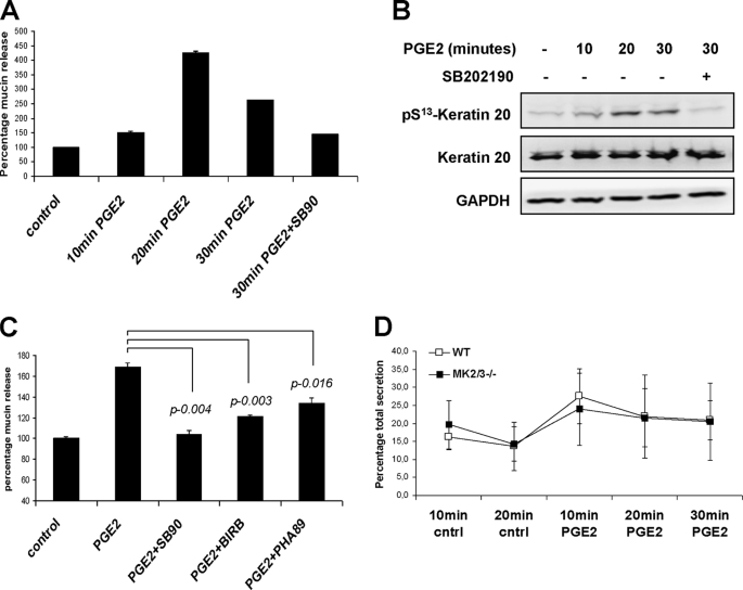
FIGURE 8.
K20 phosphorylation in PGE2-induced mucin secretion. A, differentiated HT29-MTX cells were stimulated with 5 μm dimethyl-PGE2 for indicated times, and mucin-like glycoproteins in the supernatants were quantified by ELLA as described under “Materials and Methods.” Where indicated, the cells were pretreated with 5 μm SB202190 for 30 min. B, cell lysates from the same experiment were probed with pK20 antibodies. Total K20 and GAPDH are shown as loading controls. C, mucin-like glycoprotein secretion was measured in HT29-MTX culture supernatants. Cells were left untreated or were treated with dimethyl-PGE2 (1 μm) for 30 min to allow mucin exocytosis. To study the role of p38-MK2 pathway, cells were pretreated for 30 min with 5 μm SB202190 (SB90), 1 μm BIRB-796 (BIRB), or 25 μm PHA-781089 (PHA89). D, mucin-like glycoprotein content was measured in small intestinal (ileum) effluents before (10 and 20 min) and after dimethyl-PGE2 stimulation (5 μm) (10, 20, and 30 min). The total mucin secreted in the entire assay duration (50 min) was taken as 100%, and the percentage fractions of mucin secreted at each time interval were plotted. The figure shows mean ± S.D. (error bars) of seven mice of each genotype.

