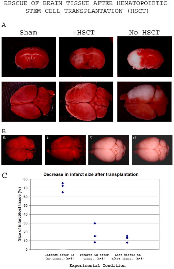
Figure 4. Rescue of brain tissue after Sca1+ bone marrow cell transplantation (HSCT).
At various times after Sca1+ cell transplantation, MCAO mice were sacrificed and their TTC-stained brains were used to evaluate the infarct.
4A-5 days after 2h transient occlusion, the infarct had a volume of 68.7% ±5.15% of the affected hemisphere (right column) and became fully white, while transplant-recipient mice (middle column) showed a clear regression of the infarct size to almost one-half to one-fourth (17.73% ±11.00%).
4B-Transplant-recipient MCAO mice surviving past 2 months after the MCAO were sacrificed at 5 months after the transplantation. TTC Staining did not reveal any infarcted region, suggesting an elimination of the dead tissue overtime. The gross morphology of the brains revealed shrinkage of the affected hemisphere, clearly noticeable in 50% of animals analyzed, as shown in panels a and b. The amount of brain tissue lost ranged from 7.76% to 15.38% of ipsilateral hemisphere.
4C-Quantification of the infarct size shown in panels 4A and 4B. The left cluster represents the infarct size in MCAO controls 5 days after 2h occlusion, the middle cluster represents the infarct size in transplant-recipient (6 million Sca1+ cells) MCAO mice 5 days after a 2h occlusion, and the right cluster represents the percentage of lost brain tissue from the ipsilateral hemisphere 5 months after Sca1+ transplantation into 2h-occlusion MCAO mice. Abbreviations: d, day; m, month; trans., transplantation

