
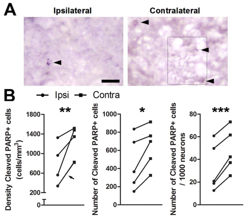
Figure 6.
T infusion near HVC prevents an increase in cleaved PARP in HVC. Photomicrographs of cells positive for cleaved PARP in HVC A) ipsilateral and B) contralateral to infusion of T. The photo is a montage of images because cells positive for cleaved PARP were in different planes of focus. Where images were spliced is indicated by a thin black line. Arrowheads indicate cells with punctate staining over the nucleus. C) Infusion of testosterone near HVC reduces the density, total number, and number/1000 neurons of cleaved PARP positive cells in HVC in birds sacrificed three days after transition to nonbreeding conditions. Arrowhead indicates measurements for two animals that share nearly identical values and thus have overlapping symbols. Asterisks indicate a significant difference across hemispheres (one-tailed paired t-test; * = p ≤ 0.05, ** = p < 0.01) Scale bar = 20μm