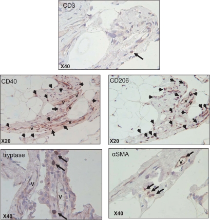
FIG. 3.
Identification of different cell types present in obese oWAT fibrosis. Serial sections of human oWAT were stained for markers of T-lymphocytes (CD3), mast cells (tryptase), fibroblastic cells (αSMA), and for CD40+ and CD206+ macrophages. The arrows show positive cells in fibrosis area revealed with DAB system (brown staining). Nucleuses were stained with hematoxylin (blue staining). V, vessel. (A high-quality digital representation of this figure is available in the online issue.)

