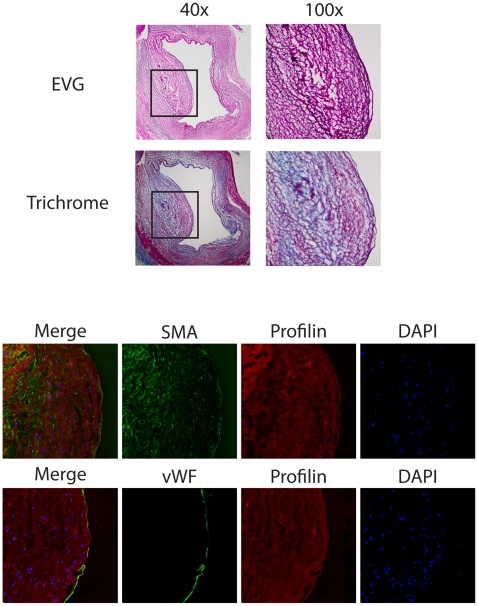
Figure 1. Expression of profilin in human atherosclerotic plaque.
Analysis of consecutive sections from a coronary artery of a representative patient with coronary artery disease. Upper panel: EvG and Masson's trichrome staining at 40×, inset was magnified at 100×. Middle and lower panels: Immunofluorescence staining for profilin (red), α-smooth muscle actin (SMA, green), and von Willebrand Factor (vWF, green). DAPI-staining (blue) was performed to visualize nuclei.

