
Figure 2. Synaptic vesicle exocytosis detected by whole-cell patch clamp recordings.
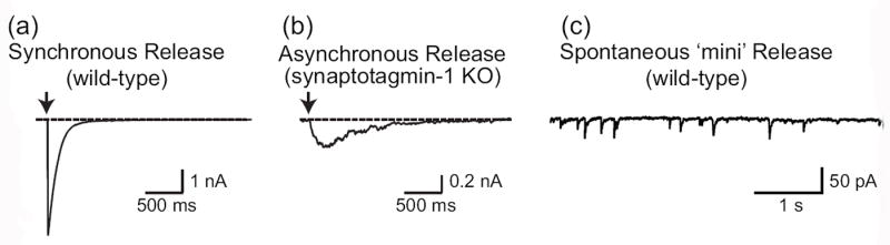
Images depict representative traces of postsynaptic currents illustrating the three different forms of synaptic exocytosis: evoked synchronous release from wild-type synapses (a), evoked asynchronous release from Syt1-deficient synapses (b) and spontaneous mini release (c). Note that asynchronous release also can be recorded in some wild-type neurons upon high-frequency stimulation.