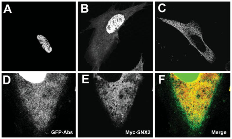
Fig. 2.

Cellular localization of GFP-Abs and Myc-SNX2. A, B: Representative images of Chinese hamster ovary (CHO) transfected with GFP-Abs. Most cells (~90%) exhibited a prominent nuclear signal (A) while some (~10%) exhibited both nuclear and cytosolic localization. C: CHO transfected with Myc-SNX2 showed a uniformly punctate distribution consistent with the endocytic compartment. D–F CHO co-transfected with GFP-Abs and Myc-SNX2 showing the cytosolic distribution of GFP-Abs (D), Myc-SNX2 (E), and merged (F) images.
