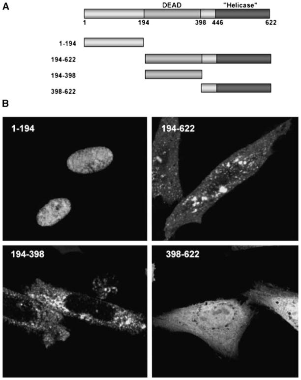
Fig. 6.
Identification of the NLS within Abs. A: Schematic diagram of full length and truncated Abs with the DEAD-box, helicase domain, and residue number as indicated. The various Abs fragments were inserted into the C-terminus of GFP. B: Representative images of transfected CHO cells. The N-terminal 194 amino acids targeted GFP to the nucleus. Deletion of this domain in Abs(194-622) and Abs(194-398) resulted in punctate cytoplasmic staining and exclusion from the nucleus. Fusion with the helicase domain in Abs(398-622) resulted in uniform nuclear and cytoplasmic staining.

