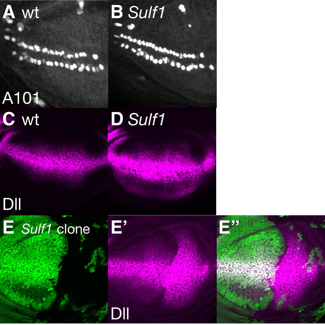
Figure 2. Wg signaling is up-regulated in Sulf1 mutants.
(A and B) neuA101 expression was visualized by anti-β-gal antibody staining of wild-type (A) and Sulf1 (B) mutant wing discs. For this and all other wing disc images, anterior is to the left and dorsal is to the top. (C and D) Anti-Dll antibody staining of wild-type (C) and Sulf1 (D) mutant wing discs. (E-E”) A wing disc baring a large Sulf1 mutant clone was stained with anti-Dll antibody. Posterior clones were induced by expression of UAS-FLP by hh-Gal4. Positions of Sulf1 mutant cells are shown by loss of GFP signal (green in E) and Dll staining is shown in magenta (E’). E” shows a merged image of E and E’.

