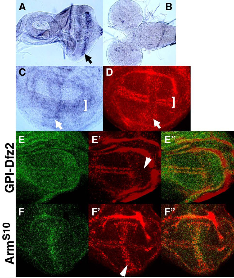
Figure 8. Regulation of Sulf1 expression by Wg signaling.
(A–D) Sulf1 mRNA expression in the developing imaginal tissues was analyzed by in situ hybridization. High levels of Sulf1 expression were detected in the morphogenetic furrow in the eye disc (arrow in A) and in specific sets of cells in the central brain (B). In the wing discs, hybridized probe was detected with a colorimetric reaction (C) or fluorescent dye (D). Sulf1 is expressed at high levels near the AP compartment boundary (arrows) and the DV border (brackets) of the wing disc. (E-F”) Sulf1 mRNA expression was examined in hh-Gal4 UAS-GFP/UAS-GPI-Dfz2 (E-E”) and dpp-GAL4 UAS-GFP/UAS-ArmS10 (F-F”) wing discs. GFP signal from UAS-GFP is shown in green (E, E”, F and F”). (E-E”) A dominant negative form of the Wg receptor, GPI-Dfz2, was expressed in the posterior compartment by hh-Gal4. Sulf1 expression was reduced by GPI-Dfz2 (red in E’). (F-F”) Expression of a constitutively active form of Arm, ArmS10, by dpp-Gal4, up-regulated Sulf1 expression (red in E’).

