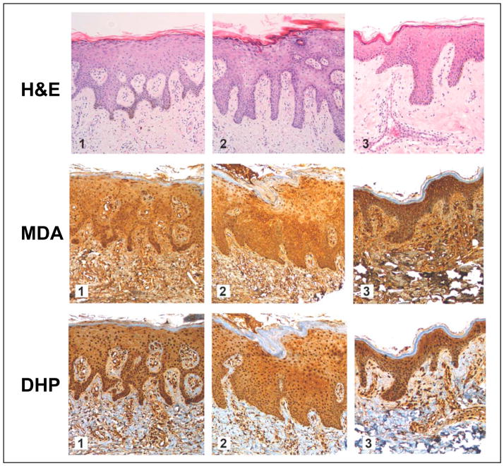
Figure 2. Immunohistochemical detection of MDA- and DHP-epitopes in healthy human skin.
A commercially available healthy human skin tissue microarray (NS21-01-TMA, Cybrdi) was processed for H&E staining (top row, specimens 1–3), pan-MDA-immunohistochemistry (middle row, specimens 1–3) using a polyclonal antibody (AP050), and DHP-immunohistochemistry (bottom row, specimens 1–3) using a monoclonal antibody (1F83). In all specimens, abundant staining for MDA- and DHP-epitopes occurs throughout the epidermal and dermal layers; Staining is most abundant in the cellular layers of the epidermis. Stratum corneum does not stain positive for either epitope. Three representative specimens are depicted.

