
Figure 1. Meta-analysis of neuroimaging data: methods and application.
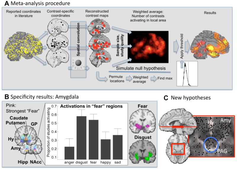
A) Coordinate-based meta-analysis. Whereas early kernel-based approaches were based on simply aggregating coordinates across a set of studies [37, 50], recent approaches explicitly test replicability across studies and allow for weighting by sample size and study quality [6, 25]. The diagram shows the procedure for one of the newer techniques, Multilevel Kernel Density Analysis [51, 52]. Reported peaks are separated by contrast map (often synonymous with study) and convolved with a spatial smoothing kernel. A weighted average map is constructed, considering sample size and other measures of study quality. The map is thresholded by randomizing the locations of the within-study activation regions many times (e.g., 10,000) and calculating the null-hypothesis distribution of the maximum across the image. This threshold provides family-wise error rate control, so that any region in the resulting thresholded map can be interpreted as more consistently activated across studies than would be expected by chance. Similar methods are available for comparing two or more task conditions (see [40]). B) Results in the sublenticular extended amygdala (Amy) from a meta-analysis comparing emotional tasks across emotion types (adapted from Table 1 in [53]). Amygdala responses are not specific for fear. C) Results in the periaqueductal gray (PAG), hypothalamus (Hy), and amygdala across studies (adapted from [51]). Replicable activation in the PAG points towards new hypothesis about PAG's previously under-appreciated role in human emotion.