Figure 1.
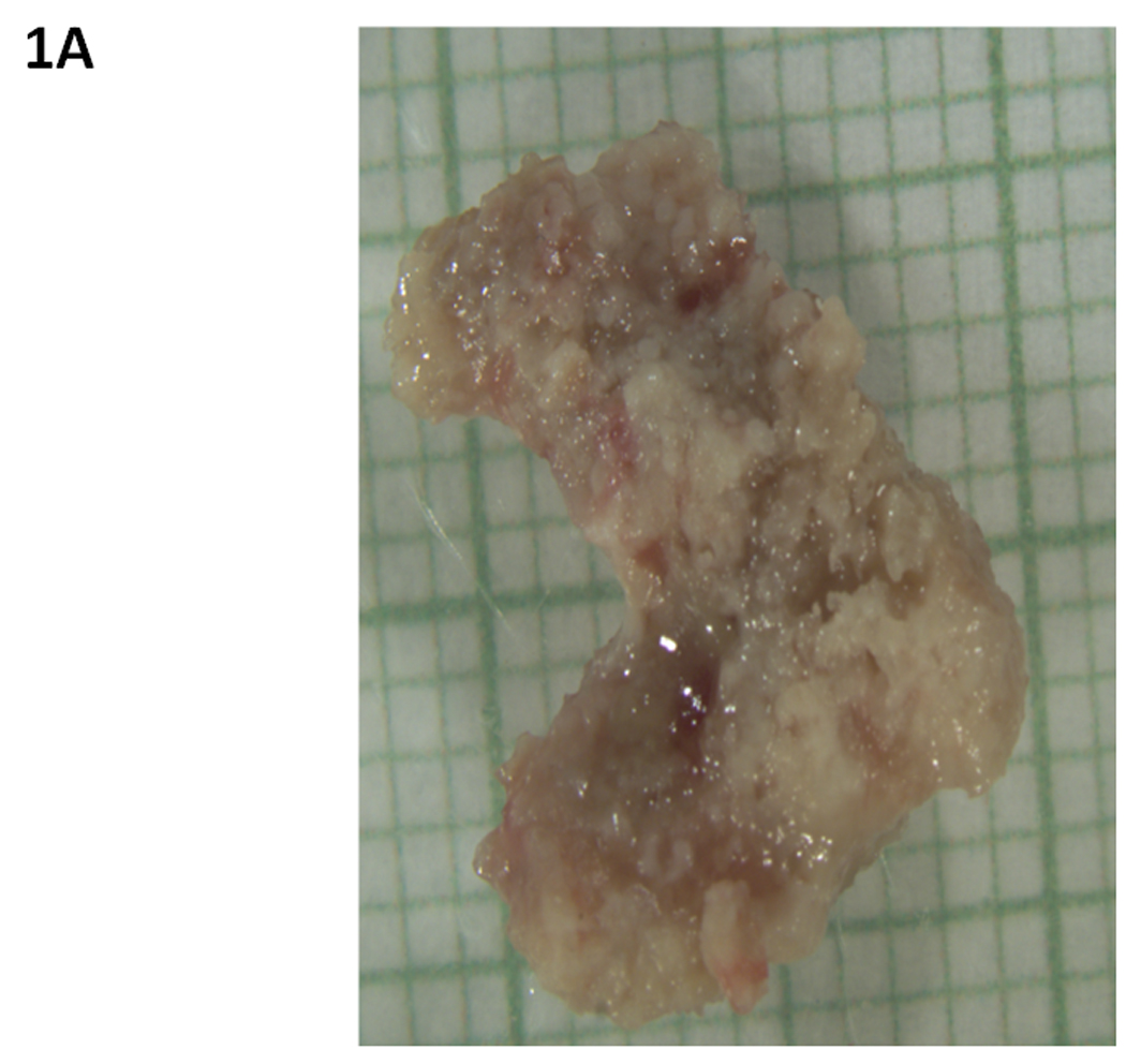
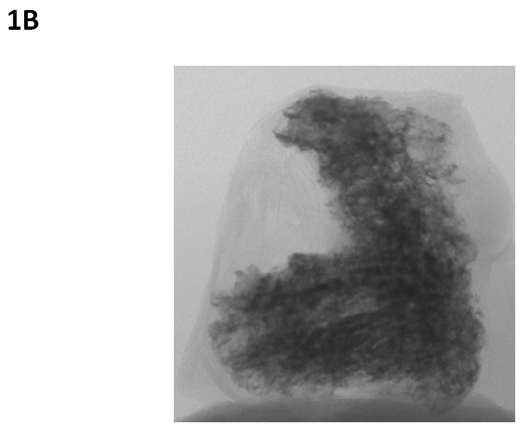
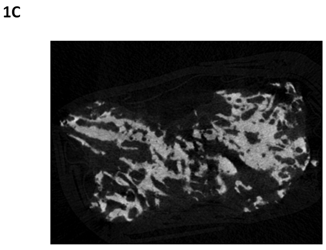
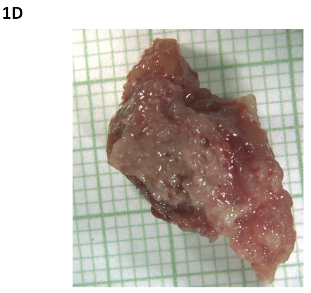
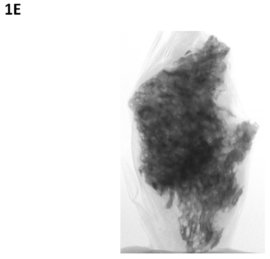
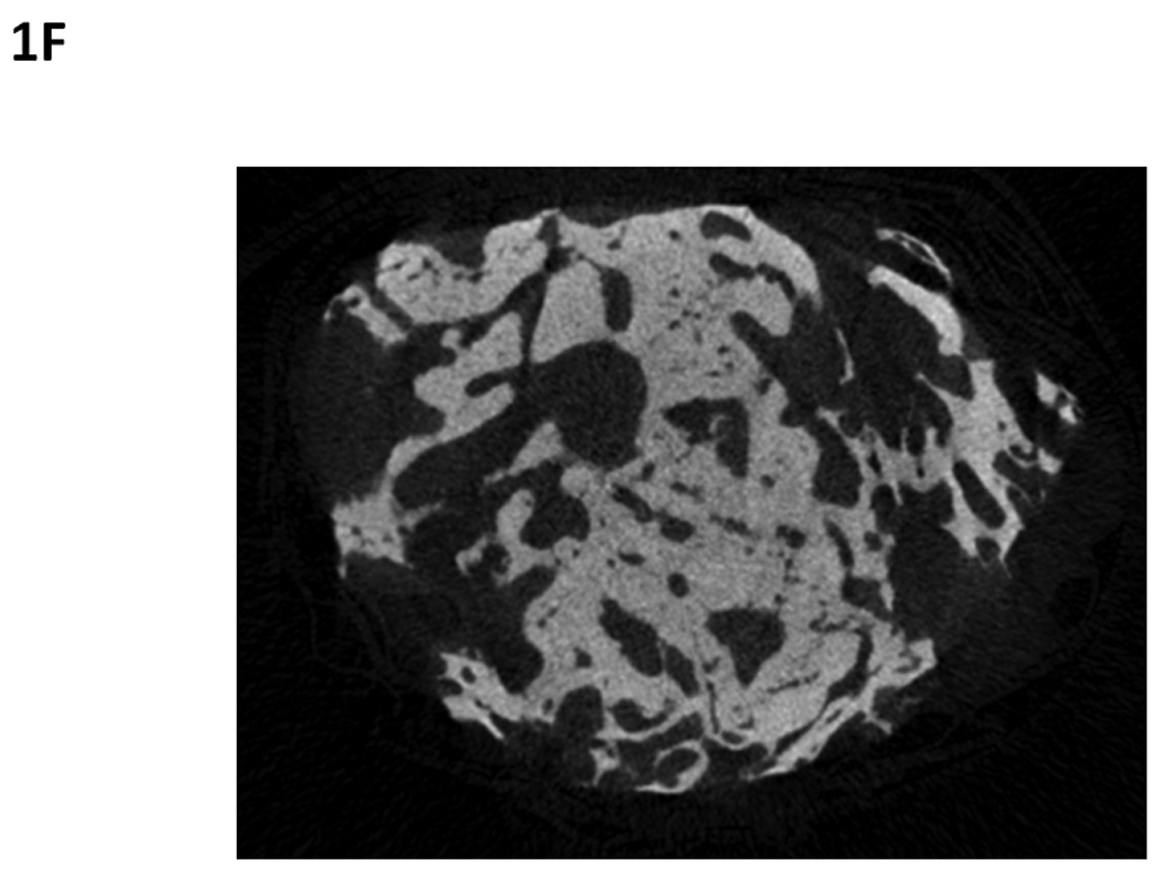
Osteonecrosis of the jaw sequestra from two different patients that had been diagnosed with ONJ for 6 months (A–C) or 48 months (D–F) prior to specimen collection. Specimens were photographed (A and D) and then scanned using micro-computed tomography which provides two dimensional projection (B and E) and cross-sectional (C and F) images. These images illustrate the similarly in the morphological structure of the specimens despite the differing durations of disease. In photographed image, each small square represents 1 mm × 1 mm.
