
Fig. 2. Cellular immune response in Hp91 immunised mice.
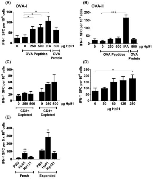
(A-B) Mice were immunised with OVA peptides in PBS (denoted as “0”), Hp91 (250 and 500 μg), or IFA. One group of mice was immunised with OVA protein and Hp91 (500 μg). Freshly-isolated splenocytes from the immunised mice were cultured in the presence of (A) OVA-I (SIINFEKL) peptide (2.5 μg ml−1) or (B) OVA-II (ISQAVHAAHAEINEAGR) (2.5 μg ml−1) in an IFN-γ ELISPOT assay. The number of IFN-γ-secreting cells was determined 18 hours later. The data shown (IFN-γ spot-forming cells per million cells) are means (+/− SEM) for 10 mice/group, except the Hp91-OVA protein group which is n=5. Asterisk, p < 0.05 between groups; Student's t-test. Data are representative of at least 3 independent experiments. (C) Freshly isolated splenocytes from PBS/OVA-peptide and Hp91/OVA-peptide immunised mice were depleted of CD8+ or CD4+ T cells and stimulated overnight in the presence of 2.5 μg ml−1 OVA-I (SIINFEKL) peptide in an IFN-γ ELISPOT assay as above. Data shown are means (+/− SEM) for 5 mice per group. (D) Mice were immunised twice with OVA-I (SIINFEKL) peptide (50ug) co-injected with 0, 30, 60, 125, or 250 μg Hp91 dissolved in PBS. Splenocytes from immunised mice were cultured in an OVA-I IFN-γ ELISPOT assay as above. Spleens were collected 6 days after the boost. Asterisk, p < 0.05 between groups; Student's t-test compared to PBS. (E) Mice were immunized with OVA peptides in PBS, Hp91 or Hp121 (250 μg). Mice received an additional boost s.c. into the contralateral flank one month after the first boost. Freshly-isolated or OVA-I-expanded splenocytes were cultured in an OVA-I IFN-γ ELISPOT assay as above. The number of IFN-γ spot-forming cells are shown as means (+/− SEM) for 4 mice per group. Asterisks, (*<0.05 and **<0.005); 2-way ANOVA.