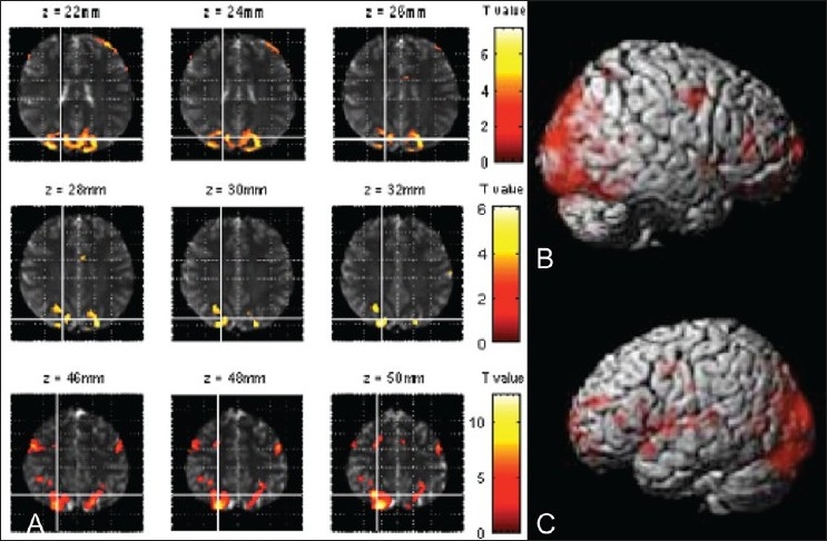
Figure 1 (A-C).

MRI images (A) show the cortical activation pattern for the whole sample for postlexical access semantic association task. Functional scans were done in a 1.5-T whole-body scanner and overlaid on an SPM anatomical template. Surface rendering of cortical activation for word concept association for all subjects for the right (B) and left (C) hemispheres (FWE-corrected, P≤0.001).
