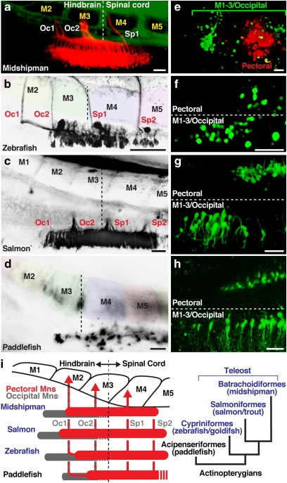
Figure 2. Embryonic alignment of pectoral and occipital motoneurons with nerves and myotomes in basal and derived actinopterygians.
(a–d) Location of pectoral motoneurons and nerves in actinopterygians revealed by lipophilic dye labelling from fin buds. The pectoral motor column began at the level of myotomes (M) 2–3 in all species studied (vertical hatching marks hindbrain–spinal boundary; also see Figure 1b,e). (e–h) Double labelling with fluorescent dextrans from fin buds and M1–3 showed that the occipital motor column began one myotomal segment anterior to pectoral motoneurons. Horizontal hatching marks midline in f–h. (i) Alignment of myotomes, nerves and motoneurons (pectoral/red and occipital/grey) with phylogenetic relationships of actinopterygians studied here (right). Paddlefish innervation pattern was deduced from juvenile gross anatomy (Supplementary Fig. S1, S2) as individual roots were not clearly visualized using retrograde labelling. All images are dorsal views with anterior to the left. Scale bars are 50 μm. Specimen stages: a (10 days postfertilization (dpf)/~5.5 mm), b (2 dpf/~3 mm), c (100 dpf/~10 mm), d (9 dpf/~13 mm), e (18 dpf/~11 mm), f (4 dpf/~4 mm), g (115 dpf/~12 mm), h (11 dpf/~16 mm).

