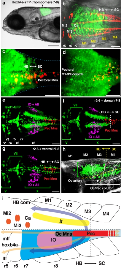
Figure 3. Embryonic alignment of precerebellar, pectoral and other hindbrain neurons in transgenic zebrafish.
(a) Rhombomere (r) 7–8 YFP expression in hoxb4a enhancer trap line. (b) Reticular (labelled from the spinal cord; red) and YFP (green) neurons showed hindbrain–spinal cord boundary between myotomes (M) 3–4. (c) Half of the pectoral column (labelled from fin bud; red) was within the hindbrain. (d) Occipital motor column (labelled from M1–3/occipital) extended to mid r8, one segment rostral to pectoral motoneurons. (e–g) Dorsal composites (e) and selected confocal planes (f, g) of pectoral (labelled from fin buds; red), inferior olive (IO) and Area II (AII) (labelled from the cerebellum; magenta) neurons in islet-GFP background (green), showing the relative position of pectoral motoneurons with major neuronal subgroups. GFP in this line is expressed in all hindbrain motoneurons, except abducens and pectoral. (h) Vagal (X) and more ventral occipital (Oc) motor columns that extended from spinal cord into the hindbrain (also see f, g). (i) Alignment of pectoral motoneurons (Pec) with other neuronal and anatomical landmarks. Pectoral motoneurons in zebrafish were located across the hindbrain–spinal cord boundary at the level of M3–5 (see Figure 2). Hindbrain motoneurons are located immediately caudal to the inferior olive, below the vagal nucleus (X) and hindbrain commissure (HB com). They are part of the occipital motor column (Oc Mns) at the level of two fibre tracts, the medial longitudinal fasciculus (mlf) and the lateral longitudinal fasciculus (llf). Other abbreviations: Mi2, Mi3 and Ca, reticulospinal neurons53. Images are dorsal (b, c, e–g, i), ventral (d) and lateral (a, h) views with anterior to the left. Scale bars are 200 μm (a), 50 μm (b–h). Specimen stages: a, c, d (4 dpf), b (2 dpf), e–h (5 dpf).

