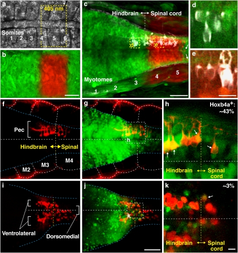
Figure 4. Origin and maintenance of pectoral motoneurons across the hindbrain–spinal cord boundary.
(a) Future hindbrain–spinal cord region in a 12 h postfertilization (hpf) embryo showing somites 1–6. (b) Transiently expressed kaede protein was photoconverted from green to red at the level of somites 4–5. Yellow dashed line in (a) marks the area of photoconversion using a laser of 405 nm. (c) The same embryo at 2 days postfertilization (dpf) showed minimal, if any, anterior–posterior migration, with labelled neurons remaining tightly clustered at the level of myotomes (M) 4–5. Arrows point to neurons that were displaced 2–3 cell diameters posteriorly. (d, e) High magnification single plane images showed photoconverted kaede to be absent in pectoral motoneurons (white) at the level of M3 (d), but present in those at the level of M4 (e). (f–k) Ontogeny of pectoral motoneurons. At 2 dpf, pectoral motoneurons were labelled with lipophilic dye from fin bud (red) and appeared as a single column at the level of M3–4 (f) where they overlapped with the hoxb4a-YFP (green) expression domain (g, shown at a higher magnification in h). At 20 dpf, pectoral motoneurons increased in number and developed into paired ventrolateral and dorsomedial columns (i). The motoneurons remained across the hindbrain–spinal cord boundary (yellow dashed line in j) demarcated by YFP expression (shown at a higher magnification in k) and exhibited a progressive downregulation of hoxb4a activity16 (arrows; h, k). Images are dorsal (a–h) and ventral (i–k) views with anterior to the left. Scale bars are 50 μm (a–c, f, g, i, j) and 10 μm (d, e, h, k). Specimen stages: a, b (12 hpf), c–h (2 dpf) and i–k (20 dpf).

